
How Autoscan helped to solve a sticky problem
The problem : Scan a slide in a battlement pattern to find all pollen grains that are fluorescent and record their positions for relocation in transmission mode microscopy.
Sounds easy : You've probably forgotten that you need to find the focal plane for each field (ie. autofocus), that you may have empty fields, that the image acquisition software has to integrate with the software for xy scanning and autofocus, and that if you are like us, you want it to all work on 32bit computer systems (Windows 98/2000/NT).
Our solution : An AS3000 stage from Autoscan Systems, a 32 bit DLL library of stage control routines from Autoscan Systems, a slow scan camera from Photometrics together with a 32bit DLL library of camera operation routines, and an image analysis package called WiT that allowed new operations to be added.
In more specific detail, the solution we came up with was to incorporate the stage control routines in the Autoscan DLL into our image analysis system by writing a new library of operators in Microsoft Visual C++ that was able to be dynamically added to WiT. This library maps the DLL routines to a form that is compatible with the imaging system, and includes such operations as "st_initialise" (stage initialisation), "st_moveto" (move the stage in xyz directions), "st_getlocation" (get the stage xyz location), and "st_status" (report stage position).
How much code is involved? Well, the average Autoscan DLL operation can be
mapped to the WiT image analysis system in about 10 to 15 lines of code. For instance
the code below is all that is required to map the stage stMoveTo32 command to a WiT
operator.
Once the AS3000 stage control and Photometrics camera controls were in place as
new operators in the WiT image analysis system, this visual programming environment
was able to be used for rapid prototyping of autofocus and scanning methods. For
instance the WiT igraph shown below allows battlement scanning to be accomplished.
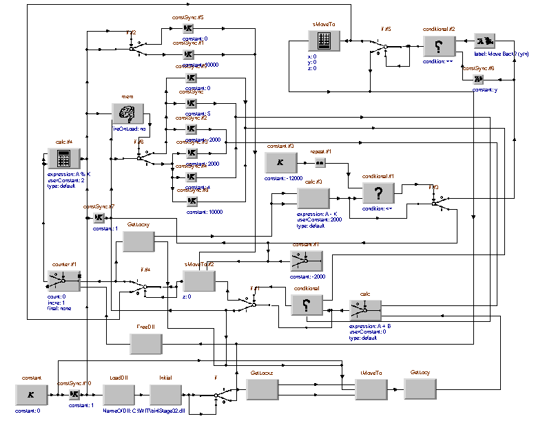
It uses the "stMoveTo" operator along with other new operators and is a visual representation of the flow of control between the various operators. This basic igraph can be used as is, or simply modified for different scan rates and step sizes by changing operator parameter values.
This scanning module is incorporated as a sub-unit of a larger igraph that controls image acquisitions and autofocus routines, with the following five principal steps:
1. Move stage to new field of view (ie. new xy location).
2. Move stage to a Z position below the specimen focal plane.
3. Acquire images and calculate focus function value until an in-focus
image is obtained.
4. Analyse to locate fluorescent objects and record x,y and z positions.
5. Move to next field of view.
The current system is capable of scanning up to 20 fields per minute for fluorescent
objects using a x10 microscope objective, with completely automatic focus finding.
This is significantly faster than can be done manually and keeps individual field
bleaching to an acceptable level.
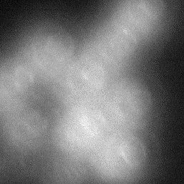
Frame 1
Frame 2
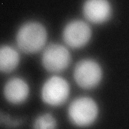
Frame 3
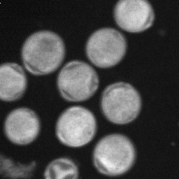
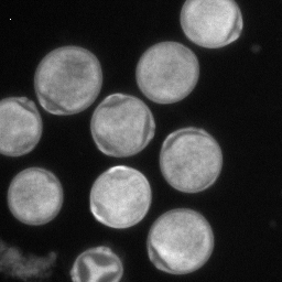
Frame 4
Focus is reached incrementally as the images above show, with frame 3 being the best focal position. Incorrect focal positioning is avoided by checking past this best focal position as shown in frame 4. This ensures the consistency of the system under conditions where objects are in different focal planes within a single image.
This combination of an advanced and adaptable image analysis system with the
Autoscan AS3000 stage has allowed us to rapidly reach our goal of an automated
scanning system for locating fluorescent pollen grains. I have no hesitation in
recommending this combination of the Autoscan AS3000 stage and the other advanced
imaging technology that we have used to other researchers who need to develop
flexible solutions, quickly that involve motorised microscopy.
Allan Jones
Research Fellow
Allergen Diagnostic Project
Institute of Respiratory Medicine
Missenden Road P.O.
PO Box M77
Camperdown NSW 2050
AUSTRALIA
Tel.: + 61 2 9351 4145
Fax : + 61 2 9351 7451
| Street Address : | Unit 56, 15 Cochranes Road, Moorabbin 3189 Victoria Australia |
| Postal Address : | PO Box 112, Ormond 3204 Victoria Australia |
| Mobile Telephone : | +61 (0)417 358 751 |
| Email : | |
| Geographic coordinates : | Lat. 37 deg. 52 min. South, Long. 145 deg. 01 min. East |
| Melway streetmap reference : | 77 K7 |
| Webmaster (Mike Krochmal VK3KRO) : | |
| NEW URL ! : | http://www.autoscan.com.au/ |